
Lower Extremity Venous Doppler
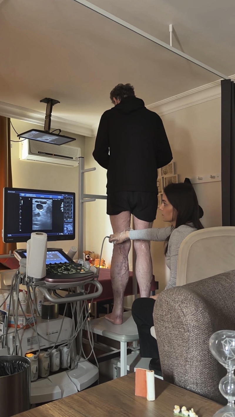
Accurate assessment of varicose vein disease requires analyzing blood flow under the right conditions, not merely visualizing the veins.
What Is a Lower Extremity Venous Doppler?
Lower extremity venous Doppler ultrasound is an examination performed to evaluate the structure of the leg veins and the blood flow within them. It is used to determine whether there is a clot, valve insufficiency, or a circulatory disorder in the veins.
What Is Assessed?
- ◆Deep and superficial veins
- ◆Direction of blood flow
- ◆Vessel patency
- ◆Presence of a clot (thrombosis)
- ◆Valve function
When Is It Performed?
- ◆Leg pain or swelling
- ◆Suspected varicose veins
- ◆Investigation of a clot (DVT)
- ◆A prolonged feeling of heaviness in the legs
How Is the Examination Performed?
The examination is carried out by assessing the vessels along their course with the ultrasound probe. When needed, the patient can be examined both lying down and standing.
At our clinic in Bakırköy, ultrasound and Doppler examinations are performed by Dr. Ahsen Bakan for patients from Istanbul, from other cities across Turkey, and from abroad.