
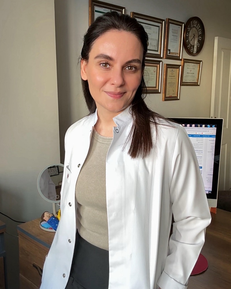
 Radiology Specialist
Uzm. Dr. Ahsen Bakan
Ultrasound is not merely about obtaining images; it is a comprehensive evaluation process that combines clinical knowledge, experience, and advanced technology.
Every examination is planned individually for the patient, and the images are assessed in detail directly by the radiology specialist.
I was born in İzmit in June 1980 and completed my primary, secondary, and high school education in Adana. Between 1999 and 2005, I studied medicine at Istanbul University, Istanbul Faculty of Medicine. In 2006, after succeeding in the national medical specialty examination (TUS), I began my residency at the Radiology Clinic of Bakırköy Dr. Sadi Konuk Training and Research Hospital, and I qualified as a Radiology Specialist in 2011.
From November 2010 to January 2011, I worked as an observer physician in Pediatric Radiology at Texas Children's Hospital in the United States.
Between 2011 and 2013, I completed my compulsory public service at Viranşehir State Hospital in Şanlıurfa. From 2013 to 2016, I served as a lecturer and radiology specialist in the Department of Radiology at Bezmialem Vakıf University.
Since 2016, I have been working intensively at my private clinic in Bakırköy, focusing in particular on thyroid, breast, pregnancy, venous Doppler, and other Doppler ultrasonography applications. I perform a wide range of ultrasound and Doppler procedures, most notably thyroid and breast biopsies, venous and arterial Doppler examinations, and carotid and scrotal Doppler ultrasonography. My core working principles are devoting sufficient time to each patient, evaluating imaging findings in detail, and offering every patient an individualized approach. I believe that ultrasound is not merely an imaging method but, when interpreted correctly, an invaluable evaluation tool that can change a patient's diagnostic and treatment course.
Special Interests and Main Areas of Practice
- ◆Thyroid ultrasonography and fine-needle aspiration biopsy
- ◆Breast ultrasonography and breast biopsy
- ◆Pregnancy ultrasonography and fetal assessment (FMF certified)
- ◆Four-dimensional (4D) color obstetric ultrasonography and fetal facial imaging
- ◆Venous Doppler and varicose vein (reflux) assessment
- ◆Carotid, arterial, renal, and scrotal Doppler ultrasonography
Certifications
My Approach to Ultrasound
An ultrasound examination is not simply about reading a report. An accurate evaluation requires examining the images live, assessing them from different angles, and supporting them with Doppler analysis when needed.
- ◆Sufficient time is allocated for each examination
- ◆The appropriate probe is selected
- ◆When necessary, the evaluation is repeated in different positions
- ◆The clinical history is interpreted together with the images
The goal is not merely to acquire images, but to contribute to an accurate clinical decision-making process.
Technology and Diagnostic Confidence
Thanks to the advanced ultrasound systems and SMI (Superb Microvascular Imaging) technology used in our center, the microvascular characteristics of tissues can be evaluated in detail.
- ◆Helps avoid unnecessary biopsies
- ◆Supports more accurate follow-up decisions
- ◆Enables reliable evaluation at an early stage
Patient Approach
Every examination is explained in a way the patient can understand. The goal is not simply to perform a test, but to provide a clear, trustworthy evaluation that eases the patient's concerns.